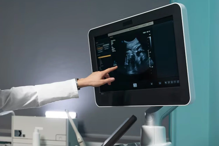
Ultrasound imaging has come a long way since its inception, with significant advancements enhancing its diagnostic capabilities. Among these, the development of 3D and 4D ultrasound imaging stands out as a transformative leap. These advanced techniques offer more detailed and dynamic views of the human body, improving the accuracy and depth of medical diagnoses and patient care.
3D Ultrasound Imaging:
-
Enhanced Visualization: Unlike traditional 2D ultrasounds, which produce flat, two-dimensional images, 3D ultrasound technology constructs a three-dimensional image of the scanned area. This provides a more comprehensive view of anatomical structures, allowing for better assessment of abnormalities.
-
Improved Diagnostics: 3D ultrasounds are particularly beneficial in obstetrics and gynecology. They offer clearer images of fetal development, enabling detailed examination of fetal anatomy, detection of congenital anomalies, and better monitoring of high-risk pregnancies.
-
Non-Invasive Precision: The technology is non-invasive and safe, using sound waves to create detailed images. This precision aids in diagnosing conditions such as tumors, cysts, and vascular anomalies with greater accuracy.
-
Patient Comfort and Engagement: The realistic images produced by 3D ultrasounds enhance patient understanding and engagement. Expectant parents, for example, can see lifelike images of their unborn child, fostering a deeper emotional connection.
4D Ultrasound Imaging:
-
Real-Time Imaging: 4D ultrasound builds upon the capabilities of 3D imaging by adding the dimension of time. This means it produces real-time, moving images, allowing for the observation of dynamic processes within the body.
-
Enhanced Fetal Monitoring: In obstetric care, 4D ultrasounds provide live-action views of the fetus, capturing movements such as kicking, stretching, and even facial expressions. This can be crucial for assessing fetal behavior and wellbeing.
-
Improved Surgical Planning: For surgeons, 4D ultrasound offers a dynamic view of organs and tissues in motion. This can aid in planning and guiding minimally invasive procedures, improving outcomes and reducing recovery times.
-
Versatile Applications: Beyond obstetrics, 4D ultrasound is valuable in cardiology for visualizing heart function in real-time, in musculoskeletal imaging to assess joint movement, and in oncology for tracking tumor growth and response to treatment.
Future Prospects:
The future of 3D and 4D ultrasound imaging holds promising developments. Advances in artificial intelligence and machine learning are expected to further enhance image clarity and diagnostic accuracy. Integration with other imaging modalities, such as MRI and CT scans, could provide even more comprehensive diagnostic information. In conclusion, the advancements in 3D and 4D ultrasound imaging have revolutionized medical diagnostics. They offer enhanced visualization, improved diagnostic accuracy, and real-time imaging capabilities that benefit both patients and healthcare providers. As technology continues to evolve, these innovations will likely play an increasingly vital role in the future of medical imaging and patient care.

